A hefty fall can leave a horse winded, but how do we know if he has suffered internal injury, asks Gil Riley MRCVS
After a heavy fall, it is important to establish what, if any, injuries the horse has sustained.
If he can’t stand, he may be winded – a spasm of the diaphragm as a result of sudden force applied to the abdomen – or have a broken limb or an injury to the spine or head. A horse who is merely winded should be back on his feet within 10 to 15 minutes. If he stays down, the likelihood is that much more serious damage has occurred.
A thorough examination should identify any architectural changes to the horse’s external anatomy. Failure to bear weight on a leg could indicate a broken bone or torn muscles in the proximal (upper) limb; moving the limb may result in crepitus, a grating sound or unpleasant sensation produced by friction between bone and cartilage in a joint or the fractured parts of a bone. Injury to the pelvic musculature or the pelvis itself can render a horse very reluctant to walk or weight-bear on one or more limbs.
{"content":"PHA+VGhlIGNoYWxsZW5nZSBpcyB0byBhc2NlcnRhaW4gd2hldGhlciBhbnkgZGFtYWdlIGlzIHN1cGVyZmljaWFsIG9yIGRlZXAsIHNvIGFueSB2ZXRzIGluIGF0dGVuZGFuY2UgbXVzdCBiZSBhbGxvd2VkIHRpbWUgYW5kIHNwYWNlIHRvIGV4YW1pbmUgdGhlIGFuaW1hbCB0aG9yb3VnaGx5LiBJZiBzZXJpb3VzIGluanVyeSBpcyBwcmVzZW50LCBpdCBjYW4gdGFrZSBjb25zaWRlcmFibGUgdGltZSB0byBnYXRoZXIgc3VmZmljaWVudCBjbGluaWNhbCBldmlkZW5jZSB0byBiZSBhYmxlIHRvIHJlYWNoIGFuIGFjY3VyYXRlIGRpYWdub3Npcy48L3A+CjxoMz5CdW1wcyBhbmQgYnJ1aXNlczwvaDM+CjxwPkZhbGxpbmcgb24gaGVhdnkgZ3JvdW5kIGNhbiBjYXVzZSBicnVpc2VzIOKAkyBydXB0dXJlcyBvZiB0aGUgc21hbGwgYmxvb2QgdmVzc2VscyAoY2FwaWxsYXJpZXMpIHVuZGVybmVhdGggdGhlIHNraW4uIElmIHRoZSBncm91bmQgaXMgaGFyZCwgZGFtYWdlIG1heSBiZSBncmVhdGVyLCB3aXRoIGFicmFzaW9uIHRvIHRoZSBza2luLjwvcD4KPHA+PGRpdiBjbGFzcz0iYWQtY29udGFpbmVyIGFkLWNvbnRhaW5lci0tbW9iaWxlIj48ZGl2IGlkPSJwb3N0LWlubGluZS0yIiBjbGFzcz0iaXBjLWFkdmVydCI+PC9kaXY+PC9kaXY+PHNlY3Rpb24gaWQ9ImVtYmVkX2NvZGUtMzEiIGNsYXNzPSJoaWRkZW4tbWQgaGlkZGVuLWxnIHMtY29udGFpbmVyIHN0aWNreS1hbmNob3IgaGlkZS13aWRnZXQtdGl0bGUgd2lkZ2V0X2VtYmVkX2NvZGUgcHJlbWl1bV9pbmxpbmVfMiI+PHNlY3Rpb24gY2xhc3M9InMtY29udGFpbmVyIGxpc3RpbmctLXNpbmdsZSBsaXN0aW5nLS1zaW5nbGUtc2hhcmV0aHJvdWdoIGltYWdlLWFzcGVjdC1sYW5kc2NhcGUgZGVmYXVsdCBzaGFyZXRocm91Z2gtYWQgc2hhcmV0aHJvdWdoLWFkLWhpZGRlbiI+DQogIDxkaXYgY2xhc3M9InMtY29udGFpbmVyX19pbm5lciI+DQogICAgPHVsPg0KICAgICAgPGxpIGlkPSJuYXRpdmUtY29udGVudC1tb2JpbGUiIGNsYXNzPSJsaXN0aW5nLWl0ZW0iPg0KICAgICAgPC9saT4NCiAgICA8L3VsPg0KICA8L2Rpdj4NCjwvc2VjdGlvbj48L3NlY3Rpb24+PC9wPgo8cD5BIGRlZXBlciBicnVpc2UgcHJlc2VudHMgYXMgYSBmbHVpZC1maWxsZWQgbHVtcCAoaGFlbWF0b21hKSwgd2hlcmUgdW5kZXJseWluZyB0aXNzdWVzIGhhdmUgYmxlZCBvciBvb3plZCBzZXJ1bSB0byBjcmVhdGUgYSDigJxiYWxsb29u4oCdIHVuZGVyIHRoZSBza2luLiBUaGUgZGFtYWdlZCBibG9vZCB2ZXNzZWxzIGJsZWVkIHVudGlsIHRoZXJlIGlzIHN1ZmZpY2llbnQgcHJlc3N1cmUgZnJvbSBmbHVpZCBhY2N1bXVsYXRpb24gd2l0aGluIHRoZSBoYWVtYXRvbWEgdG8gaGFsdCB0aGUgYmxlZWRpbmcgYW5kIGFsbG93IGNsb3R0aW5nLiBJZiB0aGVyZSBpcyBtYWpvciBkaXNydXB0aW9uIG9mIGRlZXBlciB0aXNzdWVzLCB0aGVyZSBtYXkgYmUgdG9ybiBtdXNjbGVzIGFzIHdlbGwgYXMgYnJ1aXNpbmcuIEFsd2F5cyB0cnkgdG8ga2VlcCB0aGUgaG9yc2UgY2FsbSwgc2luY2UgZXhjaXRlbWVudCB3aWxsIGluY3JlYXNlIGJsb29kIHByZXNzdXJlIGFuZCBsZWFkIHRvIG1vcmUgYmxlZWRpbmcuPC9wPgo8cD5XaGlsZSBhIGhhZW1hdG9tYSBpcyBkZXZlbG9waW5nLCBpdCBtYXkgYmUgcG9zc2libGUgdG8gY29udHJvbCB0aGUgYmxlZWRpbmcgYW5kIG1pbmltaXNlIHN3ZWxsaW5nIGJ5IGFwcGx5aW5nIGRpcmVjdCBwcmVzc3VyZSBhbmQgaWNlIHRvIHRoZSBzaXRlIOKAkyBhcyBsb25nIGFzIGl0IGlzIGluIGFuIGFjY2Vzc2libGUgYXJlYSB3aGVyZSB0aGUgaG9yc2Ugd2lsbCB0b2xlcmF0ZSBpdC4gQ29sZCBzaG91bGQgYmUgYXBwbGllZCBmb3Igbm8gbW9yZSB0aGFuIDIwIG1pbnV0ZXMgYXQgYSB0aW1lLCBhbmQgb25seSBmb3IgYXMgbG9uZyBhcyB0aGUgYXJlYSBmZWVscyB3YXJtIGFuZCBzb2Z0LiBMYXRlciwgYWZ0ZXIgdGhlIGhhZW1hdG9tYSBoYXMgc3RhYmlsaXNlZCwgd2FybSBwYWNrcyBjYW4gaW5jcmVhc2UgY2lyY3VsYXRpb24gYW5kIHN0aW11bGF0ZSBoZWFsaW5nLjwvcD4KPHA+SGFlbWF0b21hcyBhcmUgb2Z0ZW4gc3RlcmlsZSBzd2VsbGluZ3MsIHNvIHRoZXJlIGlzIGxpdHRsZSBwYWluIG9yIGluZmxhbW1hdGlvbi4gTW9zdCByZXNvbHZlLCBpZiBnaXZlbiBlbm91Z2ggdGltZSwgc28gYXJlIGJlc3QgbGVmdCBhbG9uZS4gTm8gYXR0ZW1wdCBzaG91bGQgYmUgbWFkZSB0byBkcmFpbiBhIGhhZW1hdG9tYSB1bnRpbCB0aGUgZGFtYWdlZCB2ZXNzZWxzIGhhdmUgc2VhbGVkLCBhcyBvcGVuaW5nIGl0IHVwIG1heSBhbGxvdyBibGVlZGluZyB0byByZXN1bWUuIElmIGl0IGNvbnRpbnVlcyB0byBncm93LCBob3dldmVyLCB0aGVuIHN1cmdlcnkgdG8gbGlnYXRlICh0aWUpIHRoZSB2ZXNzZWwgcmVzcG9uc2libGUgbWF5IHNvbWV0aW1lcyBiZSBuZWNlc3NhcnkuPC9wPgo8ZGl2IGNsYXNzPSJhZC1jb250YWluZXIgYWQtY29udGFpbmVyLS1tb2JpbGUiPjxkaXYgaWQ9InBvc3QtaW5saW5lLTMiIGNsYXNzPSJpcGMtYWR2ZXJ0Ij48L2Rpdj48L2Rpdj4KPHA+T25jZSBhIGhhZW1hdG9tYSBpcyDigJxtYXR1cmXigJ0sIHVzdWFsbHkgYXJvdW5kIHR3byB3ZWVrcyBhZnRlciBmb3JtaW5nLCBjYXJlZnVsIGRyYWluYWdlIHdpdGggYSBzdGVyaWxlIG5lZWRsZSBjYW4gYmUgY29uc2lkZXJlZCB0byBtaW5pbWlzZSBsb25nLXRlcm0gc2NhcnJpbmcgb3IgdG8gc3BlZWQgdXAgaGVhbGluZy48L3A+CjxwPklmIGl0IGhhcyBkZXZlbG9wZWQgYSBzdWJjdXRhbmVvdXMgcG9ja2V0LCB3aGVyZSB0aGUgc2tpbiBpcyBubyBsb25nZXIgYXR0YWNoZWQgdG8gdGhlIHVuZGVybHlpbmcgdGlzc3VlLCBpdCBtYXkgcmVmaWxsIHdpdGggc2VydW0uIFRoZXJlIGlzIGFsc28gdGhlIHJpc2sgdGhhdCBiYWN0ZXJpYSBpcyBpbnRyb2R1Y2VkLCBjb252ZXJ0aW5nIGEgc3RlcmlsZSBwb29sIG9mIHNlcnVtIGludG8gYSBwYWluZnVsIGFic2Nlc3MuPC9wPgo8ZGl2IGNsYXNzPSJhZC1jb250YWluZXIgYWQtY29udGFpbmVyLS1tb2JpbGUiPjxkaXYgaWQ9InBvc3QtaW5saW5lLTQiIGNsYXNzPSJpcGMtYWR2ZXJ0Ij48L2Rpdj48L2Rpdj4KPHA+QSBibG9vZCB0ZXN0IGhlbHBzIGlkZW50aWZ5IHRoZSBzZXZlcml0eSBvZiBhbnkgaW50ZXJuYWwgb3IgZXh0ZXJuYWwgaGFlbW9ycmhhZ2UgYnkgbWVhc3VyaW5nIHRoZSBoYWVtYXRvY3JpdCwgdGhlIGxldmVsIG9mIHJlZCBibG9vZCBjZWxscyBpbiBjaXJjdWxhdGlvbi4gUmVzdWx0cyBtdXN0IGJlIGNhcmVmdWxseSBpbnRlcnByZXRlZCwgaG93ZXZlciwgYXMgaG9yc2VzIGhhdmUgbGFyZ2UgcmVzZXJ2ZXMgb2YgYmxvb2QgaW4gdGhlIHNwbGVlbiBhbmQgbWF5IG5vdCBhcHBlYXIgb2J2aW91c2x5IGFuYWVtaWMsIGVzcGVjaWFsbHkgaW4gdGhlIGVhcmx5IHN0YWdlcyBhcyBldmVyeXRoaW5nIHN0YWJpbGlzZXMuPC9wPgo8cD5IaWdoIGxldmVscyBvZiBtdXNjbGUgZW56eW1lcyBtZWFzdXJlZCBpbiBhIGJsb29kIHRlc3QsIHByb3RlaW5zIGZvdW5kIG1haW5seSBpbiBtdXNjbGUgdGlzc3VlLCB3aWxsIGluZGljYXRlIG11c2NsZSBkYW1hZ2UuIFZlcnkgaGlnaCBsZXZlbHMgYXJlIGFzc29jaWF0ZWQgd2l0aCBicnVpc2luZyBhbmQgdHJhdW1hIHRvIHRoZSBtdXNjbGVzIG9mIHRoZSBoaW5kcXVhcnRlcnMgb3IgYmFjaywgd2hlcmUgbW9zdCBvZiB0aGUgbXVzY2xlIHRpc3N1ZSBpcyBmb3VuZC48L3A+CjxkaXYgY2xhc3M9ImFkLWNvbnRhaW5lciBhZC1jb250YWluZXItLW1vYmlsZSI+PGRpdiBpZD0icG9zdC1pbmxpbmUtNSIgY2xhc3M9ImlwYy1hZHZlcnQiPjwvZGl2PjwvZGl2Pgo8aDM+RGlmZmljdWx0IGRpYWdub3NpczwvaDM+CjxwPlJ1cHR1cmVzIG9mIGludGVybmFsIG9yZ2FucyBzdWNoIGFzIHRoZSBzcGxlZW4gb3IgaW50ZXN0aW5lLCBvciB0aGUgZGlhcGhyYWdtICh0aGUgbXVzY3VsYXIgY3VydGFpbiB0aGF0IHNlcGFyYXRlcyB0aGUgY2hlc3QgY2F2aXR5LCBvciB0aG9yYXgsIGZyb20gdGhlIGFiZG9tZW4pLCBhcmUgcmFyZS48L3A+CjxwPk1vcmUgY29tbW9ubHksIGZyYWN0dXJlIG9mIG9uZSBvciBtb3JlIHJpYnMgY2FuIGNhdXNlIHBlbmV0cmF0aW9uIG9mIHRoZSBzcGFjZSBhcm91bmQgdGhlIGx1bmdzLCBjYWxsZWQgdGhlIHBsZXVyYWwgY2F2aXR5LiBUaGUgcmVzdWx0aW5nIHBuZXVtb3Rob3JheCAoY29sbGFwc2VkIGx1bmcpLCB1c3VhbGx5IGV2aWRlbnQgYmVjYXVzZSB0aGUgaG9yc2UgaXMgc3RydWdnbGluZyBmb3IgYnJlYXRoLCBjYW4gc29tZXRpbWVzIGJlIHJlc29sdmVkIHdpdGggcmFwaWQgc3VyZ2ljYWwgaW50ZXJ2ZW50aW9uLjwvcD4KPHA+V2hpbGUgbWFueSBsaW1iIGZyYWN0dXJlcyBhcmUgdW50cmVhdGFibGUgYW5kIHJlc3VsdCBpbiBldXRoYW5hc2lhLCBzb21lIG1heSBiZSBjb3JyZWN0ZWQuIE1vc3QgY2FuIGJlIGlkZW50aWZpZWQgdXNpbmcgcmFkaW9ncmFwaHkgKFgtcmF5KSwgYnV0IG51Y2xlYXIgc2NpbnRpZ3JhcGh5IG1heSBiZSByZXF1aXJlZCB0byBkaWFnbm9zZSBoYWlybGluZSBkaXN0b3J0aW9ucyBpbiB0aGUgYm9uZSAoc3RyZXNzIGZyYWN0dXJlcyksIG9yIGZyYWN0dXJlcyB3aXRoaW4gbGFyZ2UgYW1vdW50cyBvZiBtdXNjdWxhciB0aXNzdWUgaW1wZW5ldHJhYmxlIHRvIFgtcmF5cyBvciBldmVuIHVsdHJhc291bmQgKGluIHRoZSBmZW11ciBvciBoaXAsIGZvciBleGFtcGxlLCBpbiB0aGUgbGFyZ2VyIGhvcnNlKS48L3A+CjxwPkV4YW1pbmF0aW9uIG9mIHRoZSBwZWx2aXMgaXMgZGlmZmljdWx0IGR1ZSB0byBpdHMgbXVzY3VsYXR1cmU7IHRoZSBvbmx5IHBhbHBhYmxlIGFyZWFzIGFyZSB0aGUgcG9pbnRzIG9mIHRoZSBoaXAgKHR1YmVyIGNveGFlKSBhbmQgdGhlIHR1YmVyIHNhY3JhbGUgYXQgdGhlIGhpZ2hlc3QgZXh0cmVtaXR5IG9mIHRoZSBiYWNrLiBJbXBhY3Qgd2l0aCB0aGUgZ3JvdW5kIGNhbiByZXN1bHQgaW4gYSBwaWVjZSBvZiB0aGUgdHViZXIgY294YSBicmVha2luZyBvZmYuIFNpbmNlIHRoZSB0dWJlciBjb3hhZSBzZXJ2ZSBhcyB0aGUgb3JpZ2luIG9mIHNvbWUgb2YgdGhlIGdsdXRlYWxzLCB0aGUgbGFyZ2VzdCBtdXNjbGVzIG9mIHRoZSBoaW5kcXVhcnRlcnMsIHJlY292ZXJ5IG1heSBiZSBwcm9sb25nZWQgYW5kIGxhbWVuZXNzIGxvbmcgdGVybS48L3A+CjxwPkFmdGVyIGEgZmFsbCwgc3dpbW1pbmcgY2FuIGJlIGEgdXNlZnVsIGFkZGl0aW9uIHRvIGJveCByZXN0IGFuZCB3YWxraW5nIG91dCwgYXMgaXQgYWxsb3dzIHRoZSBob3JzZSB0byByZWN1cGVyYXRlIHdpdGhvdXQgd2VpZ2h0IGJlYXJpbmcgYW5kIHRvIGFjaGlldmUgbW9iaWxpdHkgb2YgZGFtYWdlZCBtdXNjbGVzIGFuZCBqb2ludHMuIFBoeXNpb3RoZXJhcHkgY2FuIGhlbHAgcmV0dXJuIGVsYXN0aWNpdHkgdG8gdGlzc3VlcyBhbmQgbW9iaWxpc2Ugc2NhciB0aXNzdWUsIG1pbmltaXNpbmcgdGhlIGZvcm1hdGlvbiBvZiBjb25zdHJpY3Rpb25zIGFuZCBhZGhlc2lvbnMgYW5kIGJyZWFraW5nIGRvd24gdGhvc2UgYWxyZWFkeSBwcmVzZW50LjwvcD4KPGgzPlVwIGFuZCBvdmVyPC9oMz4KPHA+UmVhcmluZyBhbmQgdG9wcGxpbmcgb3ZlciBiYWNrd2FyZHMgY2FuIGNhdXNlIHNrdWxsIGZyYWN0dXJlcy4gSW4gdGhlIGNhc2Ugb2YgdGhlIGJhc2lzcGhlbm9pZCBib25lLCBhdCB0aGUgYmFzZSBvZiB0aGUgc2t1bGwsIHRoaXMgY2FuIGxlYWQgdG8gYSBjYXRhc3Ryb3BoaWMgY2VyZWJyYWwgaGFlbW9ycmhhZ2UgYW5kIHJhcGlkIGRlYXRoLiBCbG9vZCBlbWVyZ2luZyBmcm9tIHRoZSBlYXIgYWZ0ZXIgYSBiYWQgZmFsbCBpcyBpbnZhcmlhYmx5IGEgc2VyaW91cyBzaWduLCBpbmRpY2F0aW5nIGEgZnJhY3R1cmUgb2YgaW1wb3J0YW50IGJvbmVzIGluIHRoZSBza3VsbC48L3A+CjxoMz5TaXR0aW5nIGRvd248L2gzPgo8cD5JZiB0aGUgaGluZGxlZ3MgYXJlIHVuZGVybmVhdGgsIHRoZSB0dWJlciBpc2NoaWkg4oCTIHRoZSBwb2ludCBvZiB0aGUgcGVsdmlzIG5lYXJlc3QgdG8gdGhlIHRhaWwgYW5kIGFuIGFuY2hvciBmb3IgbXVzY2xlcyBpbiB0aGlzIHJlZ2lvbiDigJMgY2FuIHN1ZmZlciBicnVpc2luZyBvciBmcmFjdHVyZS4gUHJvZm91bmQgaGluZGxpbWIgbGFtZW5lc3Mgb24gdGhhdCBzaWRlIGNhbiBkZXZlbG9wIGFuZCB0aGUgbXVzY2xlcyBvZiB0aGUgdGFpbCBoZWFkIHdpbGwgc3RhcnQgdG8gYXRyb3BoeSwgb3Igd2FzdGUsIHdpdGhpbiBkYXlzLjwvcD4KPHA+TGFtZW5lc3MgcmVzdWx0aW5nIGZyb20gdHJhdW1hIHRvIHRoZSByZWdpb24gY2FuIHJlc29sdmUsIGJ1dCByZXF1aXJlcyByZWN1cGVyYXRpb24gb2YgZm91ciB0byBzaXggbW9udGhzLjwvcD4KPGgzPkZhbGxpbmcgb250byBiZWxseTwvaDM+CjxwPkZhbGxpbmcgd2l0aCBvbmUgb3IgYm90aCBoaW5kbGltYnMgc3RyZXRjaGVkIG91dCBiZWhpbmQgY2xhc3NpY2FsbHkgbGVhZHMgdG8gcnVwdHVyZSBvZiB0aGUgcGVyb25ldXMgdGVydGl1cyDigJMgYSB0ZW5kaW5vdXMgc3RydWN0dXJlIHRoYXQgcnVucyBmcm9tIHRoZSBsb3dlciBlbmQgb2YgdGhlIGZlbXVyIGFuZCBvdmVyIHRoZSBmcm9udCBvZiB0aGUgaG9jaywgaW5zZXJ0aW5nIGluIHRoZSB1cHBlciBmcm9udCBvZiB0aGUgY2Fubm9uIGJvbmUuIFRoaXMgbXVzY2xlIGVuc3VyZXMgdGhhdCBhcyB0aGUgc3RpZmxlIGZsZXhlcywgdGhlIGhvY2sgZmxleGVzIGFsc28sIHNvIGFuIGFmZmVjdGVkIGhvcnNlIG1heSBhcHBlYXIgbm9ybWFsIGF0IHdhbGsgYnV0IHdpbGwgdHJvdCB3aXRoIGEgcHJvZm91bmQgbGFtZW5lc3MuPC9wPgo8cD5GdWxsIHJlY292ZXJ5IGlzIHBvc3NpYmxlIGFmdGVyIGEgbWluaW11bSBvZiB0aHJlZSBtb250aHPigJkgYm94IHJlc3QgYW5kIHRoZSBpbnRyb2R1Y3Rpb24gb2YgYSB3YWxraW5nLW91dCBwcm9ncmFtbWUuPC9wPgo8aDM+VGhlIHNwbGl0czwvaDM+CjxkaXYgY2xhc3M9ImluamVjdGlvbiI+PC9kaXY+CjxwPlRoaXMgdHlwaWNhbGx5IGhhcHBlbnMgb24gc2xpcHBlcnkgb3IgaWN5IGdyb3VuZCAoYmVsb3cpIGFuZCBjYXVzZXMgZXh0cmVtZSBmb3JjZXMgdG8gYmUgYXBwbGllZCB0byB0aGUgbXVzY2xlcyBvbiB0aGUgaW5zaWRlIG9mIHRoZSB0aGlnaCBhbmQgbGltYi4gQ29tbW9uIGluanVyaWVzIHN1c3RhaW5lZCBhcmUgdG8gdGhlIGFkZHVjdG9yIG11c2NsZXMgb2YgdGhlIGZlbXVyIGFuZCB0aGUgY29sbGF0ZXJhbCBsaWdhbWVudHMgb2YgdGhlIGhvY2suPC9wPgo8cD5SZWNvdmVyeSBpcyBkZXBlbmRlbnQgb24gcmVzdCBhbmQgcGh5c2lvdGhlcmFweS4gV2hlcmUgbGlnYW1lbnQgZGFtYWdlIGludm9sdmVzIHRoZSBob2NrLCBhbiBhcnRocm9zY29weSAoa2V5aG9sZSkgcHJvY2VkdXJlIHRvIGRlYnJpZGUgZGFtYWdlZCBsaWdhbWVudCB0aXNzdWUgY2FuIGJlIGJlbmVmaWNpYWwgYmVmb3JlIHJlY292ZXJ5IGNhbiBiZWdpbi48L3A+CjxwPjxlbT5SZWY6IEhvcnNlICZhbXA7IEhvdW5kOyAzIERlY2VtYmVyIDIwMjA8L2VtPjwvcD4KPHA+Cg=="}
You may also be interested in:
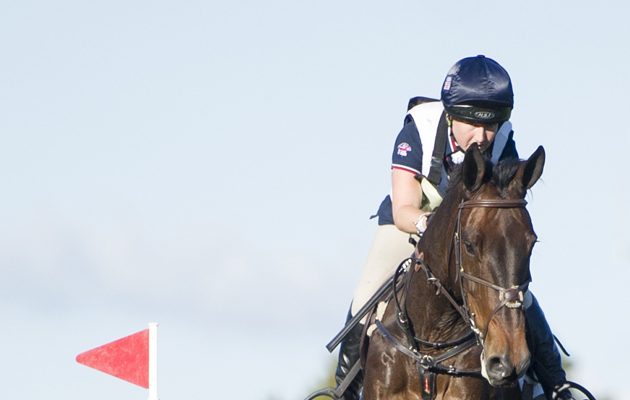
Library image.
Credit: Benjamin Clarke Photography
H&H caught up with two event riders who have sustained high profile injuries, a mental skills coach, and a soft


