When a foreign body penetrates the hoof’s horny sole or frog and goes into the sensitive tissues below, the horse’s foot has a puncture wound. It is a common cause of lameness, with the outcome varying from trivial to fatal.
A puncture wound can also be called a pricked foot, solar/hoof penetration, pus in the foot or foot abscess. Common foreign bodies include nails, sharp flints, staples, such as from fences or shavings bags, and pieces of stiff wire.
Early stages
The penetration initially causes bruising and bleeding within the sensitive parts of the hoof. Treated correctly at this stage, the lameness sorts itself out within two to three days.
If the wound becomes contaminated, however, an infection develops, with pus and gas building up within the hoof.
The pressure builds up within the hoof causing extreme pain and severe lameness. If no drainage is provided, the pus will eventually run under the sole and up the white line before bursting out at the coronary band.
Clinical signs
With puncture wounds, there are several obvious indicators:
- An initial acute lameness, which varies according to the position and depth of penetration. Often, this goes unnoticed and may subside within 24 hours.
- A more severe lameness may then develop over the next few days as infection builds up. The horse initially tries to take his weight on a certain part of the foot only – eventually, he will be unable to bear weight altogether.
- If a hind foot is affected, a stringhalt-type hopping gait may result.
- The foot feels hot and you can feel an increased digital pulse.
- The sole may be sensitive to thumb pressure, initially in a particular area, then more generally as the infection spreads.
- Often, there is a non-painful puffiness of the flexor tendon sheath above the fetlock joint.
- Eventually, pus may discharge around the coronet.
If you notice any of the above signs, you should:
- Call your vet.
- Clean and examine the foot thoroughly. If the offending item is still present, leave it there, or, if you must remove it, keep it to show your vet.
- Poultice or bandage the foot and leave the horse in a clean, dry environment.
Foot care
Your vet will thoroughly clean and examine the foot, removing the shoe if necessary. Frog injuries are particularly difficult to find, as any wounds quickly close over.
If there is nothing obvious, the vet may recommend tubbing the foot in hot water mixed with Epsom salts (magnesium sulphate), or applying a poultice. This draws the infection closer to the surface and also softens the sole, making it easier for your vet to examine.
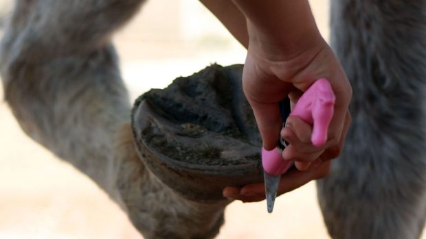
Once the site of injury has been located, the vet will dig away at the sole with a hoof knife until the pus has been released.
Frequently, the hole will be flushed with dilute hydrogen peroxide or the antibiotic metronidazole, to combat any bacteria that has found its way in.
If there is any doubt at all about the horse’s tetanus vaccination status, the vet will give him a precautionary tetanus anti-toxin.
You will need to poultice the foot twice a day for a few days. If pus has burst out at the coronet, this should be treated in a similar way.
When the discharge has ceased, the wound should be covered with a dry dressing until new horn has grown. This usually takes about a week and it is vital to keep the foot clean and dry until then. Shoeing with pads may enable a horse to be brought back into work earlier.
Antibiotics tend not to be used as they prolong lameness if drainage is inadequate. However, they are beneficial when the wound is fresh, where there is good drainage, but swelling persists, and for deep penetrations.
Complications
In most cases, the puncture wound is relatively shallow and the prognosis excellent. However, deeper wounds can be serious, with the prognosis depending on the site, direction, depth and duration of the penetration of the foreign body. (See panel above left for complications).
Other generalised complications include infections (septicaemia), laminitis and tetanus.
Continued below…
You may also be interested in:
Puncture wounds in the hoof *H&H VIP*
Deep wound treatment
The vet will need to take X-rays to determine the extent of the wound and whether there is any bony damage. If the foreign body is metallic, it can be X-rayed in-situ. Alternatively, a sterile metal probe or “contrast” material, which shows up on X-rays, can be used to delineate the wound.
In most cases, general anaesthesia, opening up the site, removing damaged, infected tissues and repeated thorough flushing is needed, plus prolonged antibiotic therapy.
Where the pedal bone is infected, all the dead, rotting bone needs to be removed, but the prognosis is generally good, providing the infection has not spread to the coffin joint or navicular bursa. Studies show that the outcome depends more on which areas, rather than how much, of the coffin bone need to be scraped away.
However, infections that tend to be associated with continuing severe lameness, often have a guarded or hopeless prognosis, particularly if the coffin joint or navicular bursa is involved.With deep wounds, the prognosis is mostly dependent on how quickly the vet begins corrective treatment.
First published in Horse & Hound magazine


