Find an equine vet
Research more about tendon and ligament injuries and MRI scans
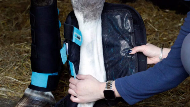
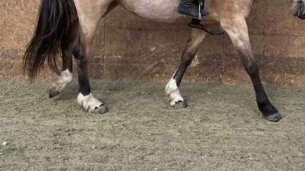
In recent years the use of magnetic resonance imaging (MRI) has greatly improved our understanding of lameness that arises from problems in the heel region of the foot.
When a lame horse came sound after a nerve block, we used to rely on X-rays to locate the source of the pain. This often led to a diagnosis of navicular.
MRI imaging has shown that a variety of problems may occur in this region, including pathology of the deep digital flexor tendon as it passes over the navicular bone and attaches to the bottom of the pedal bone.
There may be problems with the bone itself, with the ligaments of the bone or with
the ligaments of the coffin joint.
Clinicians at three American universities have pooled their records to see if they can correlate the outcome of such cases with any specific changes detected by X-ray, MRI or ultrasound scan.
They discovered that the single worst indicator for return to previous athletic ability was the presence of an injury to the deep flexor tendon.
Horses with other problems often made a complete recovery, but those with tendon damage within the back of the foot were much less likely to recover.
The use of MRI has therefore not improved the outcome for horses with pain in the heel region, but it has enabled us to make a much more accurate assessment of whether a patient is likely to make it back into work.
Read more about the latest ground-breaking advances in veterinary research on the vet clinic pages in the current issue of Horse & Hound (9 December)
Find more articles on tendon and ligament injuries and MRI scanning
Searching for an equine vet?