

When you look into your horse’s eyes, the parts you’ll notice are the light-transmitting structures — the cornea, the aqueous humour (fluid) within the anterior chamber, the lens and the gel-like “vitreous”.
In the horse, as in all mammals, this area is optically clear under normal circumstances. This clarity is a sign of ocular health, also offering the advantage that a vet can see deep into the eye using specialist instruments such as the direct ophthalmoscope.
It is possible to inspect these structures at the front of the eye, however, with little more than a small torch and cooperation from the horse.
Through the looking glass
The clear “window” into and out of the eye is called the cornea. This dome-shaped, transparent layer is an important component of the ocular focusing mechanism.
Although seemingly a simple structure, the cornea has a highly sophisticated and multi-layered anatomy that determines its optical clarity.
The healthy cornea is a relatively dehydrated structure, bathed in tears on the outside and aqueous humour on the inside. This dehydrated state is dependent upon the cellular pumps on its inner membrane, which force water out of the cornea and into the eye.
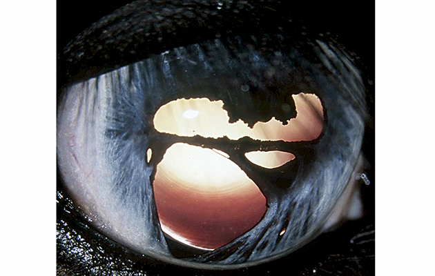
If these pumps fail, the cornea retains water and swells. The clear tissue becomes progressively opaque as its highly organised anatomy is disrupted, scattering transmitted light — rather like a sheet of clear Perspex becoming opaque along stress lines caused by bending.
These metabolic pumps are very sensitive to any injury or disease within the eye. Any opacity or greyness appearing in the cornea should prompt an owner to seek veterinary attention. Because the corneal surface is so richly supplied with nerve endings, it is one of the most sensitive structures in the body. Any surface injury will cause marked pain and an increase in tear production — another situation where immediate veterinary attention is called for.
The equine cornea appears to be susceptible to a range of diseases unique to the species, currently described using the generic term “immune-mediated keratitis”. In general, these conditions are not painful, and they typically appear as vague corneal opacities.
Causes are currently poorly understood and treatment can be challenging — and where successful, it is often lifelong. However, early recognition and treatment will increase the chances of successful control, at least, of the disease.
A lubricating layer
The thin sheet of tissue that covers the white “sclera” surrounding the cornea is called the conjunctiva. This folds around onto the inner surface of the eyelids, covering the third eyelid (the protective inner eyelid, also called the nictitating membrane).
The conjunctiva is a secretory membrane, supplying some of the important components of teardrops. It is vitally important in maintaining the health of the surface of the eye. Normally, it is a near-translucent structure, appearing slightly pink due to the underlying blood vessels. It can be heavily pigmented in some horses.
Inflammation (conjunctivitis), which in the horse is almost always secondary to some other problem in the eye, causes the conjunctiva to appear reddened and often swollen. Small tumours or growths of the conjunctiva are relatively common, particularly in eyes with little pigmentation. If caught early — which is critical — these can usually be dealt with by simple excision.
The horse’s pupil is shaped like a flattened sphere, corresponding to the visual horizon and assisting him, as a prey species, to scan this horizon continually for threats.
The dilated (open) pupil, on casual inspection, has a dark appearance. Shining a light directly along the visual axis, however, can result in a bright red/green pupil, as the light is reflected from a layer of tissue at the back of the eye called the tapetum lining. This is the reason why, at night, some animals’ eyes appear to “glow” in torchlight. Where cataracts of the lens are present, the pupil may have a pale appearance.
Pupil shape can change following injury inside the eye. Corneal pain will produce a narrow, slit-like pupil, as the muscles controlling pupil size go into spasm.
Pupil problems
The iris can be likened to the diaphragm of an old-fashioned optical camera, essentially controlling the amount of light entering the eye by altering the size of the pupil.
In most horses the iris is dark brown, but in lighter-coloured horses it may appear quite pale. In some pony breeds, or in black-coated horses, the iris can lack pigment, partially or totally. The resulting white, thin-looking appearance is often referred to as a wall eye.
These irises may also have gaps or windows in their structure, referred to as colobomas (picture, below). These have no deleterious consequences and may, in theory, permit a greater degree of depth perception in the eye. Where the iris joins the peripheral cornea, grey crescent-shaped lines may be seen. These “trabeculae” serve as drainage pathways, one of the routes through which aqueous humour leaves the eye. Clogging or collapse of this drainage angle, particularly following inflammatory disease inside the eye (uveitis), can be associated with glaucoma.
Dark-coloured, spherical structures may appear at the margin of the pupil. These are iris cysts, which arise from the inner surface of the iris and hang over its rim, attached by their umbilicus-like stalks. These are usually of no consequence, although very rarely they can grow and obstruct the pupil.
Inflammation of the iris (anterior uveitis) is usually part of a more generalised inflammatory process within the eye. When the disease is active, the iris can look muddy. The pupil is narrowed and the anterior chamber is filled with floating clumps or clouds of debris.
Affected eyes are typically very painful, but even in eyes with only minor discomfort, any cloudiness or debris within the anterior chamber is a potentially serious sign.
Iridology — the identification of more generalised disease-states from the appearance of the iris — is sometimes practised in horses. Despite having spent some 35 years studying equine eyes, this is a skill that lies well outside of both my understanding and my comfort zone.
Ref Horse & Hound; 25 August 2016