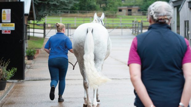
When the diagnosis of foot lameness relied upon nerve blocks and X-rays alone, conditions like navicular disease, pedal ostitis, ringbone and pyramidal disease dominated the textbooks.
Then the introduction of soft-tissue imaging using ultrasound scanners made vets realise that some of these earlier diagnoses were inaccurate and that a significant proportion of foot lameness arose from the soft tissues.
More recently, nuclear bone scanning and then magnetic resonance imaging (MRI) have opened up a world of ways in which to look inside the foot.
Both a new diagnosis and an old one have been illuminated by MRI.
Degeneration of the ligaments of the coffin joint on either side of the foot, just below the coronary band has been recognised as a specific cause of lameness.
MRI picks this up well, although a recent project by Newmarket scientists shows that the problem may be more common than even MRI reveals.
They looked at horses with apparent coffin joint ligament damage using MRI and then examined the ligaments post mortem.
Three things emerged: MRI got it right when it spotted changes in the ligaments; some lame horses had microscopic changes that did not show up on the MRI and the changes appeared to represent degenerative disease rather than traumatic injury.
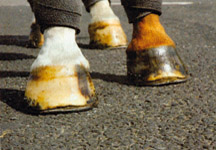
And the old diagnosis? Navicular disease, in which deep ulcers develop on the back of the bone, is an excellent candidate for MRI investigation.
Other scientists have shown the correlation between MRI images and actual disease is good.
This article first appeared in Horse & Hound magazine (27 November, ’08)