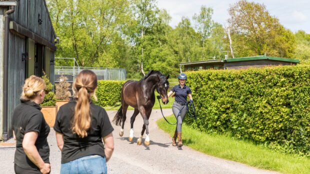
One of the most frustrating things for owners and riders is when their horse goes lame and the vet can find no good reason for it. When there are few external signs the vet has various methods to decide if the lameness is in the foot or in the shoulder.
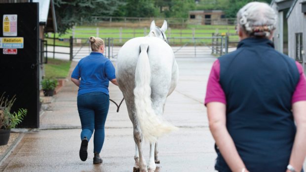
Once the horse has been trotted up and the decision on which leg is affected has been made the vet will begin by examining the whole limb by hand for evidence of pain, heat and swelling. These changes may be subtle, and any swellings are always compared with the opposite leg.
The vet will check the pulse where the arteries run down the back of the fetlock; a fast pulse may indicate pus in the foot or laminitis.
The next step is to examine the foot – is the most common cause of forelimb lameness. Hoof testers, applied firmly between the wall and the sole, will soon pick up a painful area such as an abscess or corn.
Flexion tests and nerve blocks
The vet may carry out a flexion test. This involves the vet flexing a particular joint to its full extent for a minute. The horse is then trottedup immediately after the leg is released. A few initial lame steps can be considered normal, but more might indicate a joint problem. The opposite limb is flexed for comparison.
If there are no visible or palpable signs of the cause of lameness, the vet may need to carry out a nerve block. A small volume of local anaesthetic is infused around a chosen nerve temporarily deadening it and abolishing any pain from that area.
A nerve block is usually started in the heel area, blocking off pain to the back of the foot and then, if necessary, carried on upwards until the horse shows a marked improvement, however, lameness is not always completely abolished by a nerve block.
X-rays and ultrasound
Once an area of pain is localised, it may be necessary for the vet to carry out X-rays in an attempt to look for bony damage, or an ultrasound scan, which looks for soft tissue damage. X-rays and ultrasound are only useful once the pain has been localised to one area.
Once these routine procedures have been carried out, the vet is usually close to arriving at an accurate diagnosis, but unfortunately it is not always possible to label the cause of every lameness.