A severely lame horse does not often present a vet with much of a diagnostic challenge. In contrast, horses with low-grade mild and chronic lameness can be a real nightmare to investigate. In these cases, there will probably have been a gradual onset of lameness, with the rider persevering for some time before realising that the problem is orthopaedic.
By the time the horse arrives at the clinic it is common for the case to have a long history, with copious notes and an exasperated owner, who feels that the past few months have been a stop-start episode of frustration.
So where does the vet begin? It helps if we know the horse. If it is a regular patient we will often remember how it used to move. But if we see a horse we have never set eyes on before and the owner tells us that it used to move like a dream and now has a short, stuffy action, we have to take the owner’s word for it.
The principle problem is that low-grade lameness is often bilateral — it affects both legs. This means we do not have the benefit of comparing one forelimb with the other. If both legs are affected, there will be a deterioration in the action of both. In straight lines we may observe a restricted, short, stuttering action in front or a low, straight action behind.
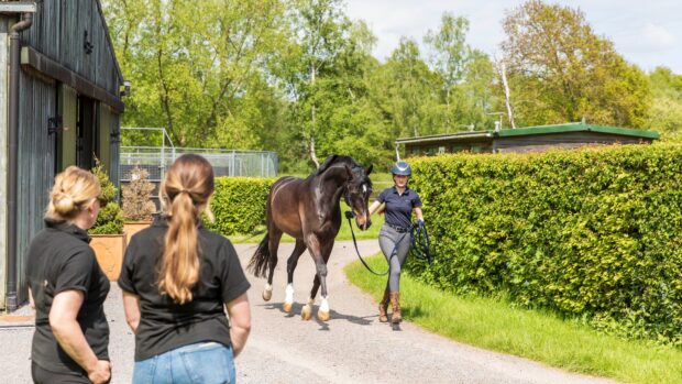
This is where circle work is invaluable because although the horse may be “sound” in straight lines, trotting on the lunge will frequently reveal lameness on whichever limb is on the inside.
We would lunge the horse on a good surface in the lungeing ring and then possibly in smaller circles on a hard surface. This provides us with a lot of clues and gives us the basis for a proper lameness work-up. In some cases there is obvious lameness in one limb, but the lungeing reveals that the problem is bilateral and that one limb is affected more than the other.
It is a myth that a horse’s movement tells the vet where the lameness is. We may have a suspicion that a certain action indicates trouble in the foot or that it may be higher in the leg, but in even the most experienced veterinary opinions, such views are at the very best only hunches and are often proved wrong.
A routine manual and digital examination of the limbs can glean further information. Swellings, heat, reaction to pressure or squeezing, flexibility of the joints, thickening of joint capsules, extra fluid in bursae or joint capsules — all these help build up a picture of the possible cause of the lameness.
Such clues are viewed as possible pointers, not as diagnostic tools. It is a big mistake to make the diagnosis of a low-grade lameness on the basis of manual examination. This is in contrast to severe lameness, in which the seat of pain can frequently be accurately localised by the vet’s hands.
The next step is to proceed to flexion tests, in which specific joints are passively flexed, held in flexion for a minute or so and the horse is then trotted away. Once again, such tests are fraught with difficulty and are useful as clues but not as hard evidence.
In the hind limb, a positive result cannot be ignored. The hock is the area most responsive to flexion tests but not the only one, so a positive response does not localise the problem to the hock. It simply indicates that a joint is involved in the lameness and that the hock is the most likely.
In the forelimb, flexion tests are very tricky because many sound horses respond slightly to flexion of the lower limb, especially as they get older. Unless the response is very marked, we tend to treat the result with some caution.
The purposes of all these exercises is to make the horse lame enough to work on. Indeed, in advance of attending the clinic for the work up of a mildly lame horse, we make certain that the patient is taken off any medication and if necessary worked to make the problem more apparent.
Once we can see a consistent lameness, we can begin localising the site of the discomfort. This is usually achieved by using regional nerve blocks — the injection of small amounts of local anaesthetic around specific nerves or into joints to deaden particular areas of the limb. After each nerve block, the horse is worked again, either in-hand or on the lunge, to see if the lameness is still present.
When we have abolished the lameness by deadening one specific area, we know that the cause lies in that region. At this stage the lameness in the opposite leg shows more clearly — because we have blocked out the discomfort in one limb, the lameness in the other becomes much more apparent.
To achieve the actual diagnosis we must wheel out the technology — X-ray machine, ultrasound scanner, bone scanner or perhaps even the keyhole camera, which requires the horse to travel to a suitable clinic and forms the next stage of the vet’s investigations.
|
||

 Get up to 19 issues FREE
 TO SUBSCRIBE