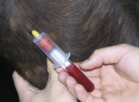
Whenever an injection is given, it carries a minute risk of causing an infection or a localised reaction. Harry, a six-year-old Thoroughbred hunter, had been with his present owners for six months when they noticed a soft, painless swelling below his left ear, which appeared temporarily while he was grazing.
Further examination revealed a slightly thickened, or “corded”, left jugular vein. We surmised that Harry had, in the past, reacted to an intravenous injection, resulting in a thickening of the jugular’s walls and a reduction in the size of the area that blood flows through (the lumen). Bloodflow back from the head to the heart on this side was therefore compromised, producing a temporary swelling (or oedema) below the ear when gravity was an additional factor. Harry appeared unaffected by all this at first, but by the autumn the oedema was increasing and the jugular vein was thickening and becoming tender.
The decision was made to refer him to ENT (ears, nose and throat) specialistG eoffrey Lane, at Bristol University, with a view to removing the offending vein. Pre-operative tests confirmed that the entire jugular was damaged and needed removing, but that there were no other abnormalities. Surgery took several hours, resulting in what the surgeon described as “the longest incision he’d ever had to make”, from jaw to shoulder with 150 to 200 stitches.
Harry made a rapid recovery and was back in work within six weeks. Scarring was negligible and the oedema has all but disappeared as his body’s bypass system has taken over. While the exact cause will always remain unknown, the outlook for a full competitive life is excellent, the only drawback being that Harry now has only one remaining jugular vein to use if he ever needs another intravenous injection.