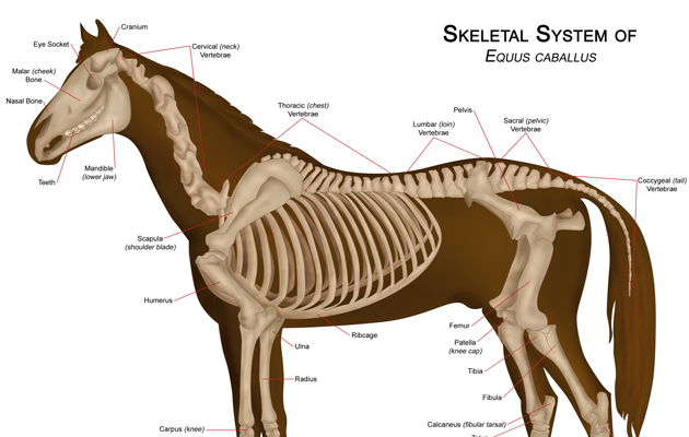
Have you ever stopped to think how a horse’s back works underneath you? It might seem complex, but if you don’t understand, you can’t train your horse correctly. These are the five sections you need to know about:
1. Cervical vertebrae
These start behind the poll and run through the middle of the neck. Muscle sits on the top.
“The reason these vertebrae are so low is because the head is heavy and horses need to be able to lift it up,” explains sports and remedial therapist Gillian Higgins of Horses Inside Out.
Most movement happens at the base of a horse’s neck. However, quite a lot of movement happens at the second vertebrae behind the skull too. This is what enables a horse to nod, and look left and right.
If the horse’s head is too high, the first vertebrae locks and they can’t flex left or right. The same happens if a horse over-flexes.
2. Thoracic vertebrae
There are 18 thoracic vertebrae, which start at the withers and connect to the ribcage. You can feel the bony projections, which are called spiny processes, at the top of the horse’s back. The first three vertebrae are hidden under the shoulder. This area also consists of muscle to enable the head to lift, plus carry its weight and that of the neck.
The spinal cord runs along the bottom of these vertebrae.
“The spinal cord is a central motorway for messages through the body to the brain and back again. It’s much lower down than most people think,” adds Gillian.
3. Lumbar vertebrae
Six lumbar vertebrae run on from the thoracic vertebrae. The lumbar vertebrae stick out to the side, as well as the top, because they do not connect to any ribs.
 “If a horse is very thin you can feel the lumbar vertebrae, but you don’t want to be able to and in most horses you can’t,” says Gillian.
There is limited rotation or flexion between the lumbar vertebrae, because nodules hold them together and reduce the range of movement. It is also very important that the saddle doesn’t finish here, because that would cause a pressure point. “The way to find the first lumbar vertebrate is to feel for the top of the last rib and run your finger up to the top. That’s it,” adds Gillian.
4. Sacrum
Next comes the sacrum. Spiny processes in the lumbar vertebrae face forward towards the head, whereas those in the sacrum face the tail. Consequently, there is a V shape where they meet. That is known as the lumbar/sacral junction, which is what enables a horse to bring its hindleg under its body.
Flexion and extension happens at this junction — that is why a riding coach refers to a horse needing to lift its back. Unless it does, the range of movement in this area is restricted and the horse cannot engage its hindleg.
Next to this is the sacroiliac joint, where the sacrum vertebrae meet the ilial at the top of the pelvis.
“There is very little movement in the sacroiliac joint, whereas the lumbar/sacral junction moves a lot. That makes this a complex area and it’s why so many problems stem from here,” explains Gillian.
5. Tail vertebrae
There are between 18 and 22 tail vertebrae, which run to the end of the dock. The spinal cord stops after the sacrum, but muscle and ligament continues to the end of the dock. This means the tail can reveal a lot about the horse’s back.
For example, a horse who is clamping its tail down or holding it to the side probably has something going on elsewhere in the back. Make sure you are familiar with how your horse naturally holds his tail — any sudden change is an early sign to call for expert advice.
Thank you to XLEquine, www.xlequine.co.uk, Scarsdale Vets Equine Practice and Horses Inside Out for their help with this feature.