Diagnosing lameness requires identification of the lame limb followed by detection of the location of pain within that limb.
Sometimes, isolation of pain is straightforward. Careful clinical examination of the limb reveals an obviously painful focus, usually associated with some degree of heat or swelling.
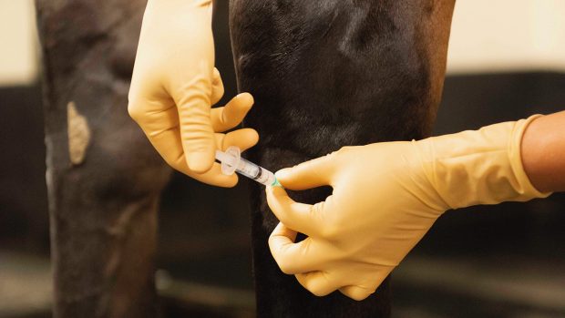
However, there are often no clues and nerve blocks are required to desensitise different areas of the limb in turn to see if there is any improvement in the degree of lameness.
Having identified the source of the pain, a variety of imaging techniques, including X-rays, ultrasound scans and, if necessary, more specialised bone scans and magnetic resonance imaging, can be used to identify the cause.
So where do problems arise? Occasionally, an obvious focus of pain is not actually the cause of lameness. There may be painful swelling in the pastern region associated with cracked heels but the source of pain causing lameness may be in the hock.
Nerve blocks are not completely reliable either. They aren’t easy to perform and require considerable practice, while the interpretation of the response is not always straightforward. They also have their limitations on where they can be successfully used on the horse’s body.
Sometimes clinical clues and veterinary technology can provide useful extra information. Areas of bone damage in higher parts of the leg may be identified using a bone scan, and major muscle tears can also be detected.
If a ligament has been damaged, abnormalities may be seen on a bone scan, where the ligament attaches to the bone. Mild damage to a deep muscle can be notoriously difficult to identify.
Obviously, lameness can only be diagnosed successfully if the correct limb is identified and investigated. This is not always as easy as it sounds, especially if there is more than one lame limb. The lameness itself can also be confusing. A head nod is usually evident with forelimb lameness, but can also be apparent with some hindlimb lamenesses.
Lameness that is very sporadic in nature can be extremely difficult to investigate. Episodic bleeding into a joint can cause transient, very severe lameness. Equally a mobile bone fragment may cause lameness that is only evident under certain circumstances.
Lameness that is only apparent when the horse is ridden can also be challenging to the vet. Sometimes, lameness that is low-grade in-hand or on the lunge is much more obvious when the horse is ridden. This can make further investigation through nerve blocks easier. However, lameness that is only apparent when the horse is ridden may be caused by the rider or by ill-fitting tack.
Lameness diagnosis requires a broad-minded but logical approach, experience and time. A detailed clinical examination, repeated if necessary, is essential to identify potential sources of pain.
If an obvious source of pain is not readily apparent, then nerve blocks are needed, but these must be interpreted in the light of the results of the clinical examination. If the pieces of the jigsaw don’t fit together, it may be necessary to start again or seek the advice of a specialist.
|
||

 Get up to 19 issues FREE
 TO SUBSCRIBE