Want to read the latest veterinary advice on identifying and treating stifle injuries? Join H&H VIP for £1 a week or less today.
Article below originally published online April 2005
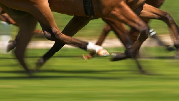
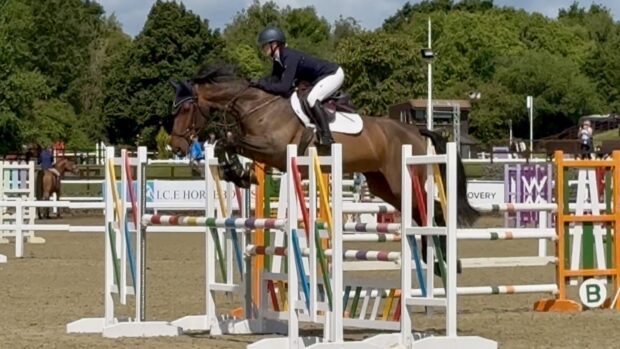
Horses jumping fixed fences at speed are particularly prone to hitting their stifles, which can result in an injury even if the front of the hind limbs has been greased.
The degree of lameness usually, but not invariably, reflects the severity of the damage. Some degree of soft tissue swelling may develop, although the absence of detectable swelling does not preclude severe damage.
If lameness is severe the stifle should be X-rayed. This is generally not an emergency and can wait for 24-48hr until the horse can be moved to a suitable facility. If lameness is mild and improves rapidly, there is probably no serious underlying injury, merely bruising. But if lameness persists the horse should be X-rayed.
A standard X-ray of the stifle comprises views from the side and back, but this may be inadequate to detect a fracture of the inside of the patella. This area can be examined by holding the leg in a shoeing position, placing the X-ray cassette under the stifle and holding the X-ray machine above it. This results in a “skyline view” of the patella. It is crucial even if bony abnormalities have been seen in other views, because a horse can sustain a fracture of more than one bone simultaneously.
The three areas most susceptible to severe injury are the tibial crest, the trochlear ridges of the femur and the patella.
Fractures of the tibial crest vary in shape and size. Sometimes there is a crack, whereas in other cases a whole piece of bone may be fractured. Depending on the precise location of the fracture, it may be prone to becoming displaced from the parent bone by the pull of the patellar ligaments.
The treatment is dependent on the size and precise location of the fracture and whether or not it is widely displaced. The vast majority of these types of fracture heal satisfactorily with rest alone.
The healing process is monitored by serial X-rays. Initially the horse is strictly confined to box rest, but when a reasonably stable fibrous union has been achieved the horse starts daily walking exercise to promote bony union of the fracture.
In the early days it may be preferable to stop the horse from lying down, because a large non-displaced fracture could become displaced by the muscle pull as it gets up. The horse can be tied via a long rope or chain and a ring, which can slide on an overhead rope extending from one side of the stable to the other. This allows the horse to walk about, but prevents it from lying down. Most horses tolerate this well.
The majority of horses are able to return to work after 3-6 months. However, if a large fracture is substantially displaced this has to be treated surgically. This involves placing a plate over the fracture using multiple screws. This usually works very well, although severely traumatised bone may be vulnerable to splitting during or after screw insertion. The prognosis for return to competition is generally good.
Fractures of one of the trochlear ridges of the femur often results in several small pieces, which must be surgically removed. This is easily performed using keyhole surgery if the pieces are immobile. It is more of a challenge if they are mobile.
During arthroscopic surgery of the femoropatellar joint, fluid runs into the joint to keep it inflated, therefore allowing a good view of the joint. If bone pieces are highly mobile they can be difficult to find and remove. Fortunately these fractures usually occur at the lower part of the trochlear ridges, a region not subject to pressure from the patella. Joint function is therefore not compromised and the prognosis is good.
Small avulsion fractures (when a fragment is torn off) may occur from the top of the patella. These generally require no other treatment than rest. Once the horse becomes sound it is generally safe to resume work, although the fragment may still be apparent on an X-ray.
More serious are the fractures that involve the inside part of the patella. Even if non-displaced, these must be removed because they never heal satisfactorily. They almost invariably involve the joint surface of the patella, and osteoarthritis is an inevitable complication if the fracture is left in situ.
Small fractures can be removed using keyhole surgery. Larger fractures may require a bigger incision, but this can be kept as small as possible by using arthroscopy to identify the location of the fracture and free the fragment from any attachments. A small incision can then be made directly over the fragment.
During the arthroscopy procedure, the entire femoropatellar joint can be inspected to ensure that there is no other damage, and any bone debris flushed out.
Generally, surgical removal of the fracture results in a favourable prognosis for return to full function, in the absence of cartilage damage. But the initial injury is sometimes more complicated. There may be associated muscle damage, or tearing of the joint capsule, or tearing of the ligamentous attachments to the patella. All these factors make the prognosis much more guarded.
- This veterinary feature was first published in Horse & Hound (24 March, ’05)