

The hock is a complex and hard-working joint, operating in conjunction with the stifle to allow the powerful muscles of the upper limb to produce rear-end propulsion. Not surprisingly, the hock region is one of the most common sites of lameness in horses from all disciplines.
The joint comprises the high-motion tarsocrural (or tibiotarsal) joint, along with three low-motion joints — the proximal intertarsal, distal intertarsal (or centrodistal) and tarsometatarsal joints.
The tarsocrural joint is where the vast majority of flexion and extension movements occur in the hock. Due to the shape of the joint surfaces, this is limited to a single plane of movement (unlike a human ankle — the equivalent joint — which not only can flex and extend but also move inwards or outwards). The range of motion of the lower hock joints is very small, in contrast, although not insignificant.
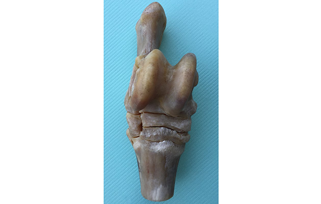
Specimen of the hock
When these small, low-motion joints develop arthritis, the normal, free-gliding functions of the joint start to fail during movement. As the problem progresses, the hock is sometimes described as “fusing”. This natural fusion is the end result of bone spavin of one or more of the low-motion hock joints. Once moderately advanced, a firm, bony swelling develops on the inside front of the hock, over the affected joint.
Bone spavin is a degenerative condition much like arthritis in humans. It can be considered a wear-and-tear condition and may be accelerated by more or harder work. Conformation and genetics are also involved in development of the problem — horses need not have a long or hard work history to suffer with the condition.
Most often, bone spavin is first identified in middle-aged horses between around nine to 11 years of age. It is not uncommon, however, to find early signs of arthritis in horses much younger than this, and some horses will develop lameness from bone spavin at only a few years of age. A wide range of breeds and disciplines are affected.
Treatment options
When considering treatments, it is important to understand that the majority of these are used to manage the condition and the associated lameness rather than trying to cure the problem. This is because the joint has become degenerative, which is irreversible.
A wide range of treatments are used for bone spavin. The most common initial approach is to inject the affected joint with an anti-inflammatory drug (a corticosteroid), similar to cortisone. Horses not being used for competition are then typically managed with long-term administration of low doses of non-steroidal anti-inflammatory drugs, such as phenylbutazone (“bute”), suxibuzone or meloxicam.

X-Ray of arthritis of the lower hock joint. The joint has narrowed and new bone has formed but it has failed to fuse naturally
Bisphosphonate treatments such as tiludronate and clodronate are also commonly used and administered by intravenous injection to alleviate bone pain.
The practice of fusion through intervention aims to eliminate movement in the low-motion joints entirely — making the hock permanently pain free. This approach is usually reserved for when medical treatments are no longer working.

A normal hock
To achieve fusion, any remaining cartilage needs to be removed from the joint so that the bone surfaces can come into contact and eventually grow together. There are various methods used to remove cartilage and to try to promote hock fusion, including laser surgery, injection of alcohol or a chemical called monoiodoacetate (MIA) into the joint, or by surgical drilling of the joint.
Pros and cons
There are pros and cons of each approach to fusing the lower hock joints. Regardless of technique, fusion takes between six and 12 months to occur.
● Surgical drilling: drilling causes fusion of the joint, at least as “spot welds” at the sites of drilling. Due to the low risk of complications and good outcomes in many cases, this is the preferred technique of joint fusion for many surgeons. Despite this, it usually takes six to nine months for the joint to fuse and for lameness to improve sufficiently for the horse to return to work.
● Injection techniques: quick improvements may be seen following alcohol injection, but this is more likely due to desensitisation than fusion
of the joint — at least initially.
A sustained improvement is not seen in all cases and outcome is inconsistent, with around 60% of cases reportedly improving to some extent long term.
Both alcohol and MIA can be injected into the standing, sedated horse, but general anaesthesia is often recommended when using MIA to minimise the risk of leakage out of the joint and damage to the skin and soft tissues. In some horses, MIA injection causes significant pain during the first day or two following treatment.
MIA reliably causes destruction of cartilage and eventual joint fusion. Occasionally, however, the two low-motion joints communicate with the upper joints. Any injected alcohol or MIA that ends up in the upper joints can cause permanent and often severe lameness.
● Laser surgery: this is performed under general anaesthesia. Similar to treatment with alcohol injection, the benefits may not be limited to fusion — some horses reportedly come sound before fusion occurs on X-rays and are able to return to work quicker than with other techniques.
Laser surgery causes smaller “spots” of fusion, however, than MIA injection or surgical drilling.
What’s the prognosis?
In the early stages of bone spavin, it is not uncommon for vets to advise an owner to put the horse on bute and keep working him in the hope that the joint will eventually fuse naturally. Although this is a good approach in many cases, the concept that “all hocks eventually fuse” is a falsehood. It is the exception rather than the rule which gets to this point naturally. Most horses will show lameness again as soon as the painkillers are stopped.
Successful fusion of the lower hock joints by intervention enables most horses to return to work and experience a good improvement in lameness. No technique is 100% successful at achieving fusion, however. Success rates for drilling are typically around 70%, with results following alcohol injections seemingly less consistent.
If bone spavin is detected at a vetting, as a purchaser you should proceed with caution.
It is of vital importance that the severity of spavin is assessed with X-rays and that veterinary advice is sought. Mild spavin changes that are not making the horse lame are a relatively low risk for development of lameness in the short or medium term (months to a couple of years), but, because the condition is degenerative and progressive, long-term lameness problems may occur.
In addition, once bone spavin has been detected on a vetting, insurance companies will exclude the hocks from cover. Any treatments that may be necessary in the future will be at the new owner’s expense.
Bog vs. bone spavins

Bog spavin
Bog spavin and bone spavin are colloquial terms describing distinctly different conditions, but due to the similar names they are often confused.
Bog spavin describes a fluidy swelling of the high-motion tarsocrural joint — the “bog” is the fluid-filled swelling you can feel, most commonly at the inside front of the hock.
A small bog spavin is a common, incidental finding of no concern, but larger bog spavins are most usually a result of bone chips in the joint.
The bony growth of a bone spavin occurs lower down the hock, frequently affecting both legs.
Ref: Horse & Hound; 16 March 2017