A keratoma is a rare, benign tumour of the keratin or horn-producing cells. These cells usually form the hard, water-resistant hoof capsule that encloses and protects the sensitive structures in the foot.
The cells that produce the wall are located at the top of the hoof wall at the coronary band. The entire wall continuously grows down from this point alone, in a similar way that the cells that produce a human’s fingernail are located under the cuticle.
The cells that produce the sole are located at the bottom of the pedal bone. Therefore, the only dividing and living cells that produce the hoof capsule are found at the coronary band and along the bottom surface of the pedal bone. The hard external hoof consists of dead cells.
If anything alters the normal pathways controlling the activity of the dividing cells, it may result in a constantly increasing mass of abnormal tissue, with a keratin core, between the outer hoof capsule and the pedal bone.
Although it is thought that chronic irritation may make the horse prone to the condition, many horses diagnosed with keratomas have no history of either infection nor injury at the site. Any size, breed or gender of horse may succumb to the problem — it may simply be bad luck.
Signs of keratoma
Some horses may remain sound, but they generally show a mild, intermittent lameness that gets gradually worse. The only indication may be an abnormality of the contour of the sole or wall noticed during routine farriery.
Keratomas of the wall tend to originate from the coronary band and can cause bulging and distortion of the wall, which is visible externally. Keratomas of the sole are generally circular and their size can vary from 0.5-2cm in diameter. In either case, widening of the white line with dense rubbery white tissue is frequently apparent.
The first signs to look for are the characteristic changes in contour of the wall and sole, and an abnormal area of the white line. X-rays can then be used to examine the mass and any damage to the underlying pedal bone. This happens if the mass has exerted sufficient pressure to cause absorption of the bone. The result is a smooth bordered semicircular defect in the bone.
Magnetic resonance imaging (MRI) has also been used to define accurately the margins of the abnormal tissue and the extent of the damage, making it easier to decide how to treat it.
Treatment
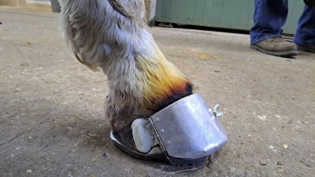
“If in doubt, get it out” is the best approach. Treatment of a keratoma is based on removal of the abnormal tissue, but is often easier said than done. The appropriate section of the outer hoof wall must be removed to allow access to the underlying keratoma.
Depending on the position and extent of the tumour, this may be achieved with the horse standing under sedation, with local anaesthetic blocks to numb the foot pain. Otherwise a general anaesthetic will be required. In either case, it is essential to ensure removal of all abnormal tissue — especially if it goes as far as the coronary band — otherwise the tumour will keep growing.
A commonly used surgical technique involves making two parallel cuts either side of the keratoma, from the sole up the wall as far as is necessary, to ensure the whole of the mass is removed. The wall is then removed by pulling it up and effectively “unpeeling” the hoof wall from the bottom of the hoof to above the tumour. The tumour can then be completely removed.
The wound is covered with a sterile dressing until it dries out. Suitable support is provided for the hoof capsule, if necessary, by means of a bar shoe. Once the wound has dried out, hoof reconstructive material can be used to protect the area while the hoof wall grows down. This can take between six and 12 months. Most horses recover well from keratomas, as long as the entire tumour is removed.
H&H 18 August 2005