
The diagnosis of navicular disease in horses has been redefined since the use of magnetic resonance imaging (MRI) to scan the feet of lame horses.
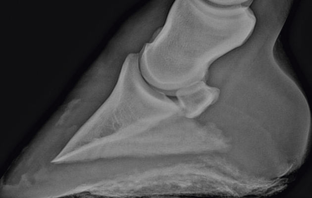
The navicular bone is the small bone at the back of the foot. The deep digital flexor tendon (DDFT) passes around this bone as it tracks down the back of the pastern and attaches to the pedal bone.
Until MRI was widely available, the two main diagnostic tools were the response to nerve blocking the back of the foot and apparent changes in the navicular bone as seen on X-rays. If a horse blocked sound to the low-volume nerve block of the heels or if X-rays showed blotches or irregularities of the navicular bone, the horse would be diagnosed as having navicular disease.
However, MRI scanning has since shown that there are several different possible problems or injuries within the hoof that will block out with traditional nerve blocking of the heels. These include damage to the DDFT or to the ligaments of the coffin joint or navicular bone, as well as damage to the bone itself.

Vets at the Animal Health Trust recently reviewed case files of more than 700 lame horses in which nerve blocks, X-rays and MRI scans were used to diagnose disease within the hoof or hooves of the forefeet.
Was there any consistent correlation between the eventual proven cause of the lameness established by MRI or postmortem exam and the changes seen on X-ray or the response to the nerve blocks?
If X-rays were of any value, it might be expected that horses suffering from true disease of the navicular bone, rather than any other structure, would have the worst X-ray findings. When all the results were analysed, however, this was not the case.
Even a careful and previously accepted scoring system for navicular bone disease based on X-rays did not match or correlate with the true cause of the lameness. And response to nerve blocks could not predict which structures inside the foot were causing pain and lameness.
This sobering result shows that although nerve blocks can confirm that the horse has foot pain, X-rays of the feet have limited value in making a precise diagnosis.
Ref: Horse & Hound; 20 August 2015