

With its fine bones, labyrinthine passages and air-filled cavities, the equine head is an anatomical wonder. Such complexity, however, can lead to difficulties when problems arise, especially those seated deep within the sinuses or the roots of the teeth.
We can now examine the equine head in ever more detail, thanks to the increasing sophistication of computed tomography (CT) scanners. Many owners are familiar with the “slice by slice” imaging created by CT, but how exactly is this technology helping vets deal with a host of head-related issues?
Lucy Meehan MRCVS, one of the few UK-based European veterinary specialists in equine diagnostic imaging, explains that the capabilities of CT go well beyond those of conventional X-ray.
“X-ray squashes three-dimensional information onto a two-dimensional image, making it hard to distinguish the finer detail,” she says. “A CT scanner is basically an X-ray machine, but its doughnut shape allows it to spin 360˚ around an area such as the head to capture information from all angles.
“This ‘lump’ of info can be sliced in any way, so we can examine complex areas,” adds Lucy. “The tissue and bone can be seen clearly without the superimposition of other structures. Computer wizardry can generate three-dimensional models from this data, allowing a vet to visualise damage and plan treatment.”
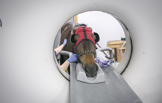
The CT scanner at Langford Veterinary Services in Bristol, where Lucy is based, is one of less than a dozen in the UK able to do head scans in the standing patient, eliminating the risk of general anaesthesia.
“The scanner can capture 16 ‘slices’ per revolution, each 1-3mm thick, and 50-70cm long,” says
Lucy. “The horse’s head needs to go through the aperture only once — in two minutes we can acquire more than 800 high-resolution images.”
Access all areas
Such a large amount of data allows virtual access to structures including the pharynx and larynx, the guttural pouches and the hyoid apparatus, facilitating investigation of dental, sinus and neurological disease.
“One of the main uses of CT is to examine teeth,” says Lucy. “We can only see the outer surfaces of the tooth on X-ray, but with CT we can slice through the head to view different cavities within the tooth.
“The live part of the tooth is called the pulp,” she adds. “Tooth infection shows up on a CT scan as gas within the 3mm-wide pulp chambers. We can see the alveolar bone that surrounds the roots of the teeth to form sockets — when these sockets enlarge, this usually means infection. We can also spot extra teeth that may not be seen orally.”
Since sinusitis can be caused by tooth root problems, CT is invaluable in investigating problems within these hard-to-access areas.
“With primary sinusitis we need to rule out dental disease,” explains Lucy. “We sometimes find a hole in a tooth socket going into the sinus.
“Headshaking is more complex, but it’s vital to rule out everything else. Many horses are headshaking due to pain from the trigeminal nerve, the main sensory nerve, so we can look at this in more detail. And we can examine the temporohyoid joint, which can cause behavioural problems and pain if diseased.
“CT is invaluable for detecting breaks in fine bones, such as those between the eyes and over the nose,” adds Lucy. “We can see displaced pieces of bone, and, if bone has been depressed, how far, and use the images to plan reconstruction.”
One shortcoming of CT is the inability to see detail in the brain.
“An MRI scan of the brain is more revealing,” Lucy says. “We can delineate the margins of a tumour with CT if we use injectable contrast, but it’s expensive and difficult to inject enough iodine-based dye.”
Standing start
The key to successful CT scanning is good preparation. The horse’s head is positioned on a table and fed through the 70cm aperture. This is achieved by loading the horse onto a moveable platform, which glides along runners using a compressed air system. While he must stand squarely, supporting himself, he should be sufficiently sedated to prevent any movement.
“We first give the horse a full clinical check, before inserting a canula into his neck to administer a small dose of long-acting sedative,” explains Lucy. “We then walk him onto the ramp to position him on the platform, giving him a reasonable dose of sedative once he’s in place.
“The machine is noisy and can be distracting, so we fit him with blinkers and put cotton wool in his ears. We also tape his ears back so they don’t catch on the scanner.”
With the horse’s head extended as far as possible, the air platform is raised — a tricky time.
“Some horses can panic, but most are fine,” adds Lucy. “We boot or bandage them for safety, plus there’s an emergency failsafe device to stop the flow of compressed air and make the platform stable.
“The radiation risk is considered minimal. There is always the chance of colic after sedation, but it’s a small risk with such a short sedation.”
The future of CT
The images are processed and assessed. Digital files can be sent to specialists worldwide in the case of unusual findings.
CT scanning has revolutionised our ability to deal with problems in the head and distal (lower) limbs, but further developments could extend its capabilities.
“One limitation is that we can only reach the first two vertebrae in the neck,” says Lucy. “The Royal Veterinary College and Liphook Equine Hospital are now using scanners with an extra-wide bore, made for bariatric [obese] humans. These are able to image the entire neck, although the horse has to be under general anaesthetic.
“Most of the equipment in equine hospitals is designed for humans and adapted for horses,” adds Lucy. “While the technology used in equine-specific CT scanners is still in its infancy, the first models are becoming available.
“Another possibility is the use of robotic arms to scan the standing horse. This could offer much better motion correction — an interesting development given that an issue with the conventional ring-shaped scanner is the horse moving.”
Ref Horse & Hound; 6 October 2016