Hindlimb lameness is more common than people realise, particularly when the onset is gradual, subtle and affects both hindlegs the same.
Often a coexisting frontleg lameness detracts from an underlying problem behind, vets find that once they have resolved a problem in front, a hindlimb asymmetry then becomes apparent.
If you suspect your horse is “not right behind” then observe him in his stable or paddock.
Does he:
If your horse exhibits any of the above then carry out the following:
If you have any concerns call your vet.
Preparing for the vet’s examination
An accurate history will help your vet enormously, and should include:
Clinical examination
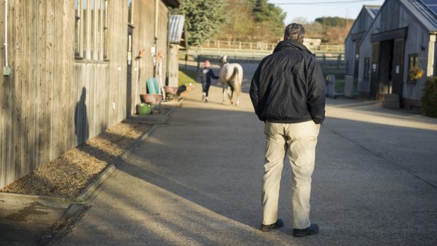
The vet will carry out, but in greater detail, the checks you have already done yourself. He will also perform flexion tests and may want to see the horse ridden under saddle.
Further tests
Some veterinary centres now use force plates and high speed treadmills to detect and evaluate subtle gait abnormalities.
Blood tests can help to differentiate true hindlimb lameness from horses whose gait abnormality is due to tying up or an infection.
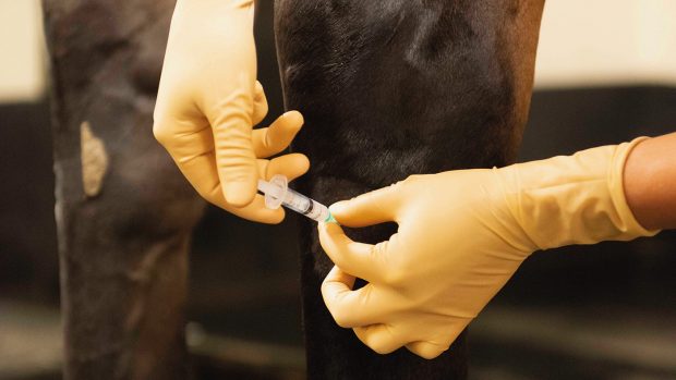
Nerve blocking anaesthetises selective areas and temporarily desensitises them. If an improvement is seen it strongly suggests the pain isin that particular area.
Intra-articular anaesthesia injects a local anaesthetic directly into a particular joint with the aim of showing a more specific area of pain.
Radiography shows up bony problems, but is really only practical for conditions affecting the stifle downwards because of the sheer bulk of the horse’s hindquarter anatomy.
Ultrasonograph: is useful for detecting soft tissue problems such as ligaments and tendons. Also conditions affectingbony surfaces, such as fractures, particuarly where radiography is impractical, as in the pelvis.
Nuclear scintigraphy or bone scanning accurately illustrates “hot spots” or bone abnormalities – in the limb.
Arthroscopy the vet to passes a telescope into the joint under anaesthesia which allows him to visualise the internal structure of that joint, including lesions.