BEFORE discussing pelvic fractures in horses, you need to understand the anatomy of horse’s pelvis, which connects the spine to the hindlimbs. Many powerful muscles attach to this structure; during locomotion, massive forces generated by the hindlimb musculature are transmitted through the pelvis into the spine, propelling the horse forward.
The equine pelvis is formed of three zones: the ilium, pubis and ischium, with the left- and right-hand sides forming a mirror image of one another. The ilium extends upwards and in front of the hip joints on either side, to form broad, curved surfaces called the ilial wings – which end at the obvious bony prominences called the tuber coxae. The pubis and ischium extend behind the hip joint and form the floor of the pelvis, connecting left and right sides at the rigid central joint called the pubic symphysis. The ischium ends with the tuber ischii, the bony prominences positioned either side of the tail.

Types of pelvic fracture in horses
THE most common type of pelvic fracture seen in horses outside racing is of the tuber coxae. Due to its superficial position on the side of the pelvis, this bony prominence takes the majority of collisions with stable doors or fences, or if a horse slips and lands on concrete, for example. These fractures may be seen with skin wounds or contusions, and the fracture severity depends on the force and angle of the collision.
Similar to these are fractures of the tuber ischii, which forms the point of the buttock. Damage is typically sustained when the horse backs into a wall, or slips with his hindlimbs beneath him to land in a sitting position.
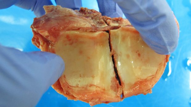
Stress fractures commonly occur within the pelvis, due to the repetitive stress of exercise. These are typically seen in racehorses, due to the repeated demands of the work performed, but also in equine athletes in other disciplines where high-intensity training is regularly undertaken.
Bone is constantly adapting, or remodelling, to ensure that it is strong enough to cope with the forces experienced during exercise. The way this material adapts without becoming too heavy is by a coupled process, where new bone production and resorption – the natural breaking down of old bone – work hand in hand.
Excessive repetitive stress can result in this balance being lost, however, which in turn can lead to a stress fracture. Stress fractures in the pelvis, like those of other bones, develop in predictable locations, the most common of which is within the ilial wing.
Signs of pelvic fractures in horses
SIGNS that a horse may have fractured his pelvis include moderate to severe lameness, usually evident at a walk. Some types of pelvic fracture may also lead to more specific changes in how the horse walks. If the tuber ischii is fractured, for example, the horse may be reluctant to bring the affected hindleg forward as this will tighten the muscles running over this area.
Swelling of the affected region may also be seen. This can range from obvious bleeding and distortion to more subtle signs of asymmetry. A crunching feeling, termed crepitus, is sometimes evident upon examination as the bone fragments rub against each other. Most often, any associated bleeding is self-limiting, but if one of the major vessels – for example, those running across the shaft of the ilium – is damaged, a fatal internal bleed can occur.
Diagnosis can often be made from the clinical features described. Sometimes, more information is gained by internal examination via the rectum. Attempts at diagnosing pelvic fractures with X-rays are often unrewarding, because the huge muscles surrounding most of the pelvis reduce what can be seen.
Ultrasound, while not often used to diagnose fractures, is very useful for pelvic fractures in the horse. Only the surface of the bone is visible, but any displacement of the fracture fragments or significant bleeding into the muscle can typically be seen.
Stress fractures, in particular, are difficult to identify, and can easily be missed or misdiagnosed as a “pulled muscle”. But it is important to diagnose a stress fracture as continued exercise may result in a much more severe or even life-threatening fracture.
Diagnosis often requires advanced imaging with nuclear scintigraphy (bone scan), which is a sensitive method of pinpointing the site of injury. A radioactive element called technetium-99m is injected into the area and binds to a molecule, which then attaches to the calcium-containing crystals where new bone is being formed. Radiation is emitted from this compound and an image of the bone activity can be seen with a special detector.

During a bone scan at the clinic, the camera is angled to get the best view of the ilial wing.
Treatment of pelvic fractures in horses
IN the majority of cases, treatment of pelvic fractures in horses involves conservative management. Depending on the severity and location of the fracture, and the horse’s comfort level, he will typically be given a period of stable rest ranging from six weeks to three months, before walking exercise can be re-introduced.
When there is significant risk of the fracture displacing further and causing injury to important vessels, cross-ties are often used. This involves keeping the horse tied up in the stable, wearing a padded headcollar, to prevent him from lying down and incurring further injury. If this is necessary, the horse will need close monitoring and pain management. He must be allowed regular breaks to lower his head so that mucus can drain from his nostrils, to prevent lung infection.
Surgery is rarely used to treat pelvic fracture. The requirement for a lengthy period of general anaesthesia usually rules out surgical repair in adult horses, but this has been successful in foals.
Surgical intervention is usually only required to remove problematic fragments of bone that are painful or infected, or those involved with an open wound. Ilial wing fractures, for example, often produce a spike of bone that presses against the skin and requires removal.
The severity of a fracture often relates to whether or not the horse can bear weight, despite the injury. Involvement of the hip joint or disruption of the overall stability of the pelvis are key determinants in the prognosis. The pelvis is also close to several large and important vessels, which can be damaged during fracture.
Injuries to areas that are important for muscle attachment may not impact as much on weight-bearing, and are generally less severe. A common example of this is fracture of the tuber coxae. A horse with this injury is typically very lame for one to two days, before displaying a rapid improvement in comfort levels.
This injury is often referred to as a “knocked-down hip”, as the fractured tuber coxae ends up lower than normal on the affected side. Horses who sustain this fracture generally make a full return to athletic function, despite a permanent change to the shape of the pelvis.
Meet the vets
MATT SMITH MRCVS, a Royal College of Veterinary Surgeons (RCVS) specialist in equine surgery, is a surgeon and partner at Newmarket Equine Hospital (NEH). Matt Chesworth MRCVS, also based at NEH, is a European specialist in equine surgery. This referral hospital is one of the largest in Europe, treating a wide range of leisure and sport horses from throughout the UK. Contact: 01638 782020, newmarketequinehospital.com
You might also be interested in:

A guide to box rest — how to navigate this testing time

Innovative surgery on equine fracture helps prevent catastrophic leg injury
The modern practice allows surgery to be performed without the need of general anaesthetic

Diagnosing hairline fractures with scintigraphy
Horse & Hound explains why scintigraphy or bone scanning can be one of the best ways of diagnosing a hairline

The Horse & Hound Podcast 51: Ginny Elliot | Bone scans | Wellington CDI | News round-up

Subscribe to Horse & Hound this spring for great savings

Nuclear scintigraphy (bone scans): what you need to know about this diagnostic test
Scintigraphy or bone scanning is a sensitive diagnostic tool, which can help identify the 'hot spots' of lameness
Horse & Hound magazine, out every Thursday, is packed with all the latest news and reports, as well as interviews, specials, nostalgia, vet and training advice. Find how you can enjoy the magazine delivered to your door every week, plus options to upgrade your subscription to access our online service that brings you breaking news and reports as well as other benefits.