

Heart murmurs are relatively common in horses.
The term “murmur” refers to any noise in addition to the normal heart sounds. Some of these are normal physiological flow murmurs, which occur due to the sheer size and power of the equine heart. More serious, however, are murmurs caused by a leaky valve.
It is sometimes possible to differentiate between harmless flow murmurs and those due to leaking valves by listening to the heart with a traditional stethoscope. Often, however, further testing, by a heart scan or an ECG (electrocardiogram), or both, is required to diagnose the significance of a murmur and its exact cause.
If the murmur is found to be valve-related, these tests can provide a valuable insight into the severity of the leak. Further evaluation will then be necessary to ascertain whether the horse is safe to ride, or whether he may be dangerously prone to collapsing during exercise.
ECG or scan?
An ECG measures the electrical activity of the heart — the timings of the beats and the rhythm of the organ as it pumps.
The normal rhythm of the heart is made up of different components. These different waves are called “P”, “QRS complex” and “T” waves — all of which together make up one heartbeat.
In medical terminology, a heart scan is termed an echocardiogram (often shortened to “echo”). Essentially, this is an ultrasound examination of the structure of
the heart.
Ultrasound of the heart is much more complex than it is in other areas of the horse, such as a leg tendon. There’s the challenge of working with the intricate three-dimensional anatomy, and, of course, the heart is in perpetual motion.
Ideally, an echocardiogram should be performed by a Royal College of Veterinary Surgeons (RCVS), European or American internal medicine or cardiology specialist.
The equipment capable of performing an echocardiogram uses sophisticated technologies such as “colour flow Doppler” and “pulsed wave Doppler” to evaluate blood flow and leaky valves, and to measure the speed of jets of blood that are leaking.
Because these machines tend to be big, scans are usually taken within a hospital or clinic — although a number of specialists do have mobile equipment.
An ECG and a scan are often complementary and can be used in tandem to evaluate different properties of the heart. These tests may be performed to investigate:
• heart murmur
• rhythm abnormality
• poor performance
• collapse
How are the tests done?
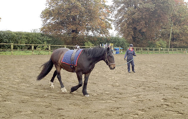
If an ECG is to be performed at exercise, three or four patches of the horse’s coat will be clipped — either within the saddle and girth area, or just to the side so that it will be out of the way if he is to be ridden. The clipped patches are cleaned and ECG pads are then stuck onto the skin, just as would happen with a human undergoing the same test.
For an echo, the hair behind the elbows on both sides is clipped. This is usually necessary unless the coat is exceptionally fine, because hair stops ultrasound and
the cardiac probe needs to have good contact with the skin. The skin is then cleaned and some ultrasound gel is applied.
The scan is usually performed with the horse in stocks. The ultrasonographer must apply relatively firm pressure to achieve good contact; if the horse is not restrained, he has a tendency to move away from the machine.
Ideally, the scan must be completed without the horse having received any sedative drugs, so that a true evaluation of the heart can be undertaken. If sedative drugs have to be used, their effect on blood pressure and therefore heart size and rate must be
taken into account.
New technology
Veterinary smartphone ECG apps have been on the market for a few years now and have proved useful for initial evaluation of a horse with an abnormal heart rhythm at the yard or an event.
If an abnormality is found, the horse can then be referred for more thorough investigation with a more detailed ECG machine. Apps have proved particularly handy for use on the racecourse, immediately post-racing, and at sport horse events such as eventing and endurance.
A number of electronic stethoscopes have been launched in the past few years that may, in time, replace the traditional stethoscope. The big bonus for the vet is being able to record the heart murmur and visualise it on a phonogram (a graphic record of sound). This allows objective documentation to be kept on the horse’s clinical record as to the shape and timing of the heart murmur.
Rider safety is paramount — we all know that riding can be dangerous, even with a fit, healthy horse. Modern cardiology techniques allow us to predict better which horses are likely to collapse when being ridden, and to allow horses with sometimes severe cardiac disease to continue to compete and do the jobs they enjoy.
Ref: Horse & Hound; 21 July 2016