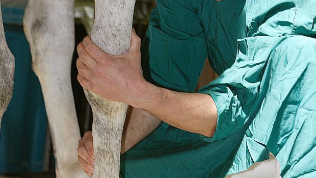
The pastern region is particularly vulnerable to the effects of direct trauma, such as a penetrating injury, due to its closeness to the ground and the lack of a protective wall like the hoof has.
The pastern has important anatomical structures close under the skin, which are susceptible to penetrating injury, while tendon and ligament structures on the back of the pasture are liable to strains.
The pastern comprises:
- The long pastern bone
- The short pastern bone
- The joint between the two bones
- Collateral ligaments which support the joint and prevent excessive rotation
- The common digital extensor tendon, which runs down the front of the pastern
- The superficial digital flexor tendon, which runs down the back of the pastern and branches into two, running either side of the deep digital flexor tendon, and inserting on to the top of the short pastern
- The deep digital flexor tendon
- The sesamoidean ligaments, which lie under the flexor tendons, running from the sesamoid bones and attaching to the pastern bones
Ringbone
The name ringbone is used to describe a variety of conditions that result in new bone formation in the pastern region. In some horses, it is used to describe the new bone formation around the pastern joint associated with osteoarthritis.
This is a condition that can only be diagnosed with certainty through X-rays. It cannot be reliably diagnosed by feeling the leg.
Many horses have small bony spurs on the top of the short pastern that are not associated with lameness. If such spurs are seen in a lame horse, it is important to inject local anaesthetic solution into the joint and assess whether lameness is relieved. If the spurs are significant, there should be a marked improvement.
If there is extensive new bone formation around the pastern joint, detectable using X-rays, narrowing of the joint space, or destructive changes in the bone underlying the joint, then it is likely that the pastern joint is the source of pain.
Low-grade osteoarthritis can often be managed successfully by treating the joint with a variety of medicines, but advanced osteoarthritis usually results in long-term lameness.
New bone formation unrelated to the pastern joint may develop as the result of direct trauma. This usually results in lameness while the bone is forming, but it should resolve, although a bony lump will persist.
Fractures
Fractures of the long or short pastern are less common in mature riding horses, than in immature Thoroughbred racehorses. Short partial fractures of the top of the long pastern occur in horses from any discipline and can present a diagnostic challenge, requiring high-quality X-rays and sometimes a bone scan. If recognised early, these fractures usually respond well to box rest and most horses are able to return to work.
Occasionally, a horse suffers multiple fractures to either the long or short pastern bone, usually the result of a major misstep. These fractures result in severe lameness and obvious instability of the lower leg — the pastern region feels like a bag of marbles. Fortunately, this is an uncommon injury, because the outcome for athletic function is poor to hopeless.
|
||

 Get up to 19 issues FREE
 TO SUBSCRIBE